Summary
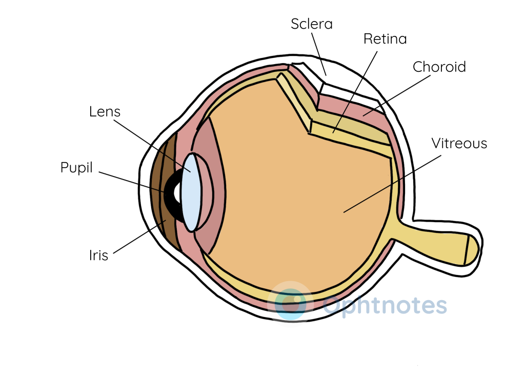
This article provides an overview of the major components of the eye, including the sclera, conjunctiva, cornea, iris, lens, choroid, vitreous, and retina. It explains the functions of each part, such as the sclera’s role in eye movement, the cornea’s refractive power, the iris’s control of light entry through the pupil, and the retina’s function in light detection. Understanding these basic anatomical features is essential for grasping the pathophysiology of eye conditions.
The sclera
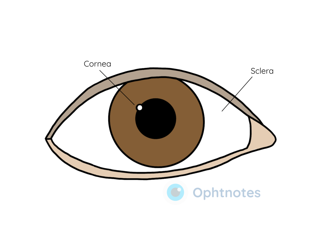
This is the white part of the eye, visible to the naked eye. It is formed of opaque tissue and is attached to extraocular muscles which control the movement of the eye.

The conjunctiva
A thin layer of transparent connective tissue covers the front part of the sclera and folds back on itself into the inside part of the eyelids. This layer of connective tissue is known as the conjunctiva.
The cornea
The dome-shaped transparent part of the eye is called the cornea. The cornea provides most of the refractive power of the eye through which light beams are focused.
The tears which line the surface of the cornea provide a medium for oxygen exchange, since the cornea itself is avascular.
Layers of the cornea
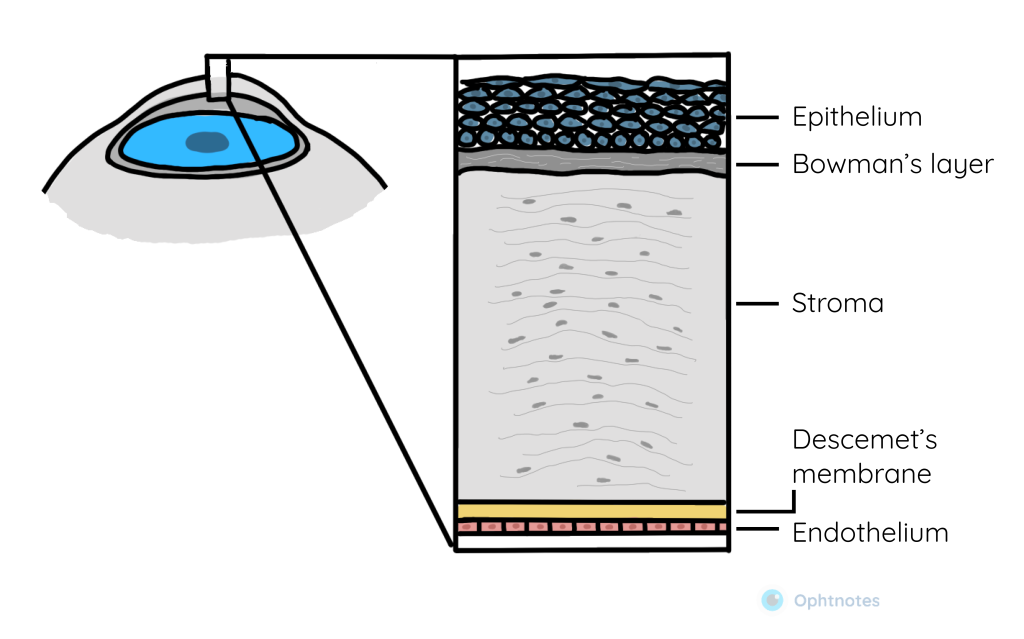
The cornea has five layers:
- Epithelium
- Bowman membrane
- Stroma
- Descemet membrane
- Endothelium
Each of these five layers have separate functions, however, we will not be covering those in this basic anatomy article.

The iris
Through the cornea, we can see the coloured part of the eye, called the iris. The iris forms an aperture through which light beams can travel into the eye, known as the pupil.

The iris also contains muscles that control the size of the pupil. The iris will constrict the pupil upon exposure to bright light and will dilate in exposure to darkness.
The lens
Once light beams pass through the pupil, they pass through a transparent globular structure inside the eye, known as the lens.
The lens can change shape to focus on objects at different distances. This is known as accommodation.
The lens is attached to the ciliary muscle through zonular fibres. The ciliary muscle contracts and relaxes to control the shape of the eye. It also produces a liquid called aqueous humour.
The choroid
This is a vascular layer between the sclera and the retina. Along with the ciliary body and iris, these three structures make up the uveal tract or the uvea.
The vitreous
Behind the lens is a gel-like substance called the vitreous. The vitreous functions to maintain the shape of the eye. It is mainly composed of water and so allows light to pass through to the back of the eye on the retina.
The retina
The inside layer of the eye where light is detected is called the retina. This is where light beams are focused onto. The part of the retina that handles central vision is called the macula and medial to the macula is the optic disc, where the optic nerve enters the back of the eye.