Summary
Optical Coherence Tomography (OCT) is a non-invasive imaging technique that enables high-resolution visualisation of retinal structures. It is pivotal in the diagnosis and management of various ocular conditions, including macular degeneration, glaucoma, and diabetic retinopathy. This article aims to provide an overview of OCT and its clinical applications.
Background
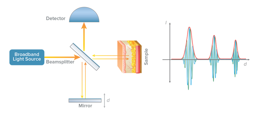
OCT operates by employing light waves to acquire cross-sectional images of the retina, providing detailed information about its layers. This technology allows for imaging that previously required invasive procedures, making it a cornerstone in ophthalmic diagnostics.
- Diagram/Image: [Insert a diagram illustrating the principle of OCT, showing how light waves are used to create cross-sectional images of the retina.]

What is OCT used for?
OCT is the primary investigation utilised to assess the structural integrity of the retina. It is particularly useful for:
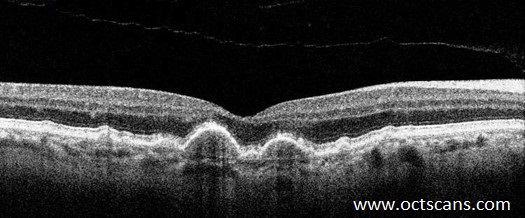
- Detecting early signs of macular degeneration
- Monitoring retinal fluid levels post-surgery
- Evaluating the thickness of the retinal nerve fibre layer in glaucoma assessment

OCT to diagnose ocular conditions
In the context of radiology, OCT serves as a vital tool for diagnosing and managing ocular conditions. It complements traditional imaging modalities such as fundus photography and fluorescein angiography by providing detailed cross-sectional views of the retina.
For instance:
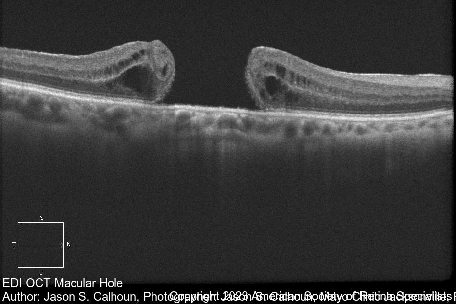
- In cases of macular holes, OCT can reveal the extent of vitreous traction and assist in surgical planning.
- In wet macular degeneration, OCT can identify the presence of fluid and abnormal blood vessels, guiding treatment decisions such as the use of anti-VEGF injections.
- Diagram/Image: [Insert an OCT scan showing a macular hole and its implications for surgical management.]

Management of conditions identified by OCT
The management of conditions identified by OCT varies based on the diagnosis:
- Macular Degeneration: Anti-VEGF injections (e.g., Aflibercept, Ranibizumab). If macular degeneration is left untreated, it could result in permanent visual loss.
- Glaucoma: Topical medications like prostaglandin analogues (e.g., Latanoprost) to reduce intraocular pressure. If left untreated, glaucoma could progress to irreversible optic nerve damage.
- Surgical Interventions: For conditions such as epiretinal membranes or macular holes, vitrectomy may be indicated.
NHS screening
With early detection via OCT, many ocular conditions have improved prognoses. The NHS Diabetic Eye Screening Programme in England will offer Optical Coherence Tomography (OCT) scans to patients in the digital surveillance pathway. This will start in October 2024 and be rolled out nationwide by October 2025.